Skip to content


Researchers have developed an AI-driven cell tracking system capable of monitoring individual cells throughout their entire life cycle — from growth and division to reproduction and dormancy — addressing a long-standing limitation in live cell microscopy.
By combining generative artificial intelligence with time-lapse imaging, the system enables continuous observation of cellular behaviour rather than isolated image snapshots. The advancement expands analytical capabilities in research laboratories and enhances single-cell analysis workflows.
The project, led by investigators at North Carolina State University, integrates algorithm development with experimental microscopy systems to improve both data interpretation and imaging efficiency.
Integrating AI with live cell microscopy
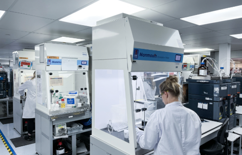
Live cell microscopy allows scientists to observe living cells in real time, but automated tracking remains challenging because cells change size, shape and position — especially under stress or during replication.
The team developed an algorithm called FIEST (Frame Interpolation Enhanced Single-cell Tracking), which applies AI-based image processing to follow cells even through significant morphological changes. Initially tested in yeast, the system was later adapted for bacteria, cancer cells and human organoids, demonstrating broad research applicability.
According to project lead Orlando Arguello-Miranda, the integration of AI has dramatically accelerated biological analysis.
Benefits for laboratory workflows
AI cell tracking offers several operational advantages:
- Continuous monitoring across generations
- Automated growth and behaviour quantification
- Reduced manual analysis time
- Improved tracking accuracy
- Scalability for high-content imaging datasets
In one example, the algorithm successfully tracked 632 cells within a complex dataset where previous tools failed — highlighting improved robustness and reliability.
Beyond academic research
High-content microscopy experiments often generate thousands of images, creating analysis bottlenecks that delay results. AI-driven tools could help laboratories process datasets more efficiently while improving data consistency.
The system is also being adapted for agricultural and environmental applications, including microscopy-based pathogen detection. Unlike DNA tests, AI-based imaging can assess organism viability by identifying distinctive morphological features.
Future outlook
Researchers are expanding AI-enabled imaging to study intracellular protein dynamics and molecular interactions within living cells. For laboratory leaders, advances in AI microscopy may influence imaging infrastructure, automation strategies and collaboration between computational and biological teams.
As AI integration progresses, laboratories could gain deeper, dynamic insights into cellular systems — moving beyond static measurements toward real-time biological understanding.
Read the full story here.