Skip to content


Researchers at King’s College London’s Centre for Ultrastructural Imaging have collaborated with Linkam Scientific Instruments to optimise a cryo-correlative light and electron microscopy stage for cryo-FIB and cryo-TEM applications. The partnership has already contributed to breakthrough imaging of the malaria-causing parasite Plasmodium falciparum, and points to a broader future for cryo-EM as the go-to technique in biological imaging.
Advanced imaging technologies are essential in life sciences research, giving detailed insights into the structure and function of organs, tissues, cells, and sub-cellular molecules and, ultimately, helping accelerate scientific research. Cryo-electron microscopy (cryo-EM) is set to transform the field of biological imaging, facilitating new discoveries in disease diagnosis and monitoring, drug discovery and development, and environmental analysis.
For researchers focusing on biological imaging, cryo-EM holds the key to unlocking more targeted analyses, allowing researchers to not only determine the structures of single molecules, but also image entire cells in their native environment. Ongoing innovation and industry collaboration in cryo-EM instrumentation is vital to support pharmaceutical and biopharmaceutical labs in harnessing new scientific discoveries and shortening drug discovery cycles.
Why cryo-EM?
In 2017 Jacques Dubochet, Joachim Frank and Richard Henderson were awarded the Nobel Prize in Chemistry for developing high-resolution cryo-EM for the structural determination of biomolecules. Before this, X-ray crystallography and nuclear magnetic resonance (NMR) were the standard technologies used for biological imaging, but both have limitations: for example, X-ray crystallography can only be carried out on crystallisable samples, while larger molecules can be difficult to distinguish using NMR.
The Nobel Prize award marked the emergence of cryo-EM as an effective and versatile alternative. By freezing biological samples to below -195 °C and then imaging them from multiple angles using an electron microscope, images can be overlaid to construct a 3D model of the sample at close to atomic resolution. Cryo-EM also forms the basis of several other techniques, including cryo-transmission electron microscopy (TEM), cryo-scanning electron microscopy (SEM) and cryo-focused ion beam-SEM (cryo-FIB-SEM). An additional benefit of cryo-EM is its ability to provide images of molecular structures in their native states, allowing researchers to acquire a more in-depth understanding of their samples. Unlike NMR, cryo-EM can easily handle larger molecules and, compared to X-ray crystallography, molecules do not need to be crystallised ahead of imaging – a significant time and cost saving. As both vendors and researchers continue to innovate with cryo-EM, it is fast becoming the go-to technique for biological imaging.
Collaboration to improve cryo-EM capabilities
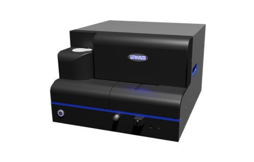
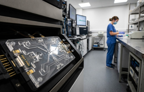
The Centre for Ultrastructural Imaging (CUI), headed up by Prof. Roland Fleck at King’s College London, UK, is the central EM facility for the university that supports both internal and external collaborators across fields including medicine, chemistry and biological sciences. The team has collaborated with Linkam Scientific Instruments to optimise a cryo-correlative light and electron microscopy (cryo-CLEM) stage to support cryo-FIB and cryo-TEM applications, addressing the needs of researchers for more targeted analysis by offering improvements in the user interface and sample transfer capability.
Cryo-TEM employs vitrification to solve near-atomic-resolution structures of a sample in close to its living state. The CUI’s cryo-TEM system, which utilises the new cryo-CLEM stage, has played an important role in the study of Plasmodium falciparum, a parasite that causes severe and often fatal malaria in, providing high-resolution images of P. falciparum red blood cells during invasion, infection and egress. Thisbreakthrough has helped further researchers understanding of how the parasite behaves, offering greater insight into treatment and management options.
For cryo-FIB analysis, the team has been using the stage to prepare lamella, and also been working on developing cryo-volume imaging for sectioning inside a cryo-FIB system, using the cryo-CLEM stage to help target cells. The cryo-CLEM stage has allowed the CUI to preserve the sample’s native environment whilst allowing for more targeted analysis of biological materials.
In addition to targeted analysis, cryo-FIB volume imaging performed using the cryo-CLEM stage opens up the possibility for observing larger structures than previously possible. Vitrified samples undergo serial milling and imaging to produce a detailed 3D reconstruction of the cell’s internal structure, revealing organelles, membranes and molecular details at nanoscale resolution. Using cryo-FIB, researchers can study cells in their near-native state and bridge the gap between molecular details and entire cellular context.
Future applications of cryo-EM in the lab
The future looks promising for cryo-EM, given the significant advancements the technique has already been able to achieve in biological imaging. Looking ahead, research institutes are now exploring the suitability of cryo-electron tomography (cryo-ET), which facilitates molecular imaging within a cell or membrane and allows researchers to view the sample much closer to its native state. Emerging technologies such as this, in addition to ongoing developments, are expediting biological imaging research timelines and lamella production. Vendor and user collaboration is also vital in supporting these developments, combining the technical expertise of vendors with the requirements of users in the lab and opening the door for further developments and streamlined methodologies in the lab.
Read more about the cryo-CLEM stage.